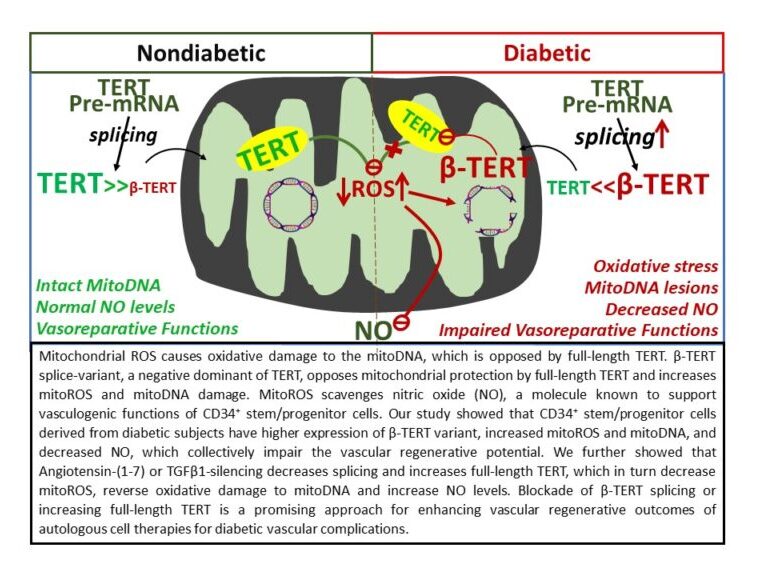
Angiotensin (Ang)-(1-7) stimulates vasoprotective functions of diabetic (DB) CD34+ hematopoietic stem/progenitor cells partly by decreasing reactive oxygen species (ROS), increasing nitric oxide (NO) levels and decreasing TGFβ1 secretion. Telomerase reverse transcriptase (TERT) translocates to mitochondria and regulates ROS generation. Alternative splicing of TERT results in variants α-, β- and α-β-TERT, which may oppose functions of full-length (FL) TERT. This study tested if the protective functions of Ang-(1-7) or TGFβ1-silencing are mediated by mitoTERT and that diabetes decreases FL-TERT expression by inducing splicing. CD34+ cells were isolated from the peripheral blood mononuclear cells of nondiabetic (ND, n = 68) or DB (n = 74) subjects. NO and mitoROS levels were evaluated by flow cytometry. TERT splice variants and mitoDNA-lesions were characterized by qPCR. TRAP assay was used for telomerase activity. Decoy peptide was used to block mitochondrial translocation (mitoXTERT). TERT inhibitor or mitoXTERT prevented the effects of Ang-(1-7) on NO or mitoROS levels in DB-CD34+ cells. FL-TERT expression and telomerase activity were lower and mitoDNA-lesions were higher in DB cells compared to ND and were reversed by Ang-(1-7) or TGFβ1-silencing. The prevalence of TERT splice variants, with predominant β-TERT expression, was higher and the expression of FL-TERT was lower in DB cells (n = 25) compared to ND (n = 30). Ang-(1-7) or TGFβ1-silencing decreased TERT-splicing and increased FL-TERT. Blocking of β-splicing increased FL-TERT and protected mitoDNA in DB-cells. The findings suggest that diabetes induces TERT-splicing in CD34+ cells and that β-TERT splice variant largely contributes to the mitoDNA oxidative damage.

